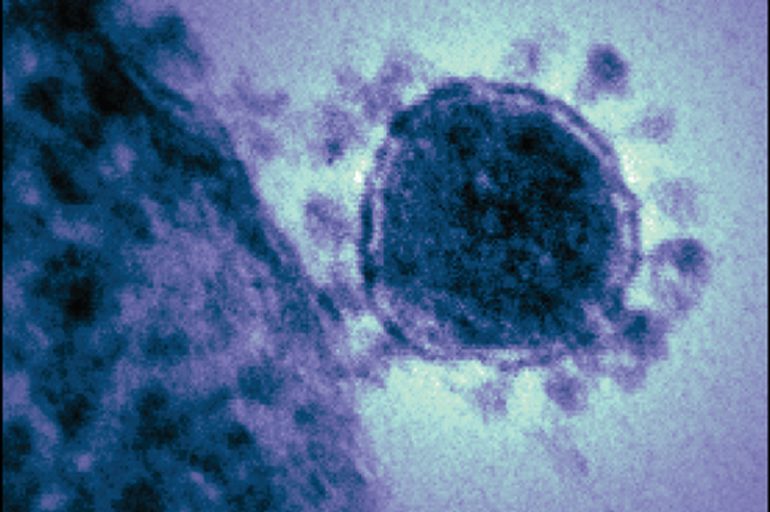
A UF virologist assisted a team of medical researchers at the Icahn School of Medicine at Mount Sinai in New York City with interpreting microscopic images of tissue samples from COVID-19 victims. The researchers found extensive damage to small blood vessels, and they propose a mechanism linking vessel injury with biological pathways that lead to an immune system in overdrive.
New research might explain the mechanism behind a constellation of odd COVID-19 symptoms, such as strokes in young people, delirium, purple toes and children with Kawasaki-like vascular inflammation.
A University of Florida research professor and virology expert, John Lednicky, contributed to a new study that supports growing evidence of COVID-19 being more than a viral respiratory illness. It’s also an infectious disease of the vascular system, the research team says.
While several recent studies point to COVID-19 as a disease that affects multiple systems, few have offered explanations for how the disease produces its signature immune system overresponse. But the new study proposes a mechanism that links damage to blood vessels with signaling pathways that lead to hyperinflammation and, in the most severe cases, a dangerous immune system shift into overdrive. This turn can happen even when patients seem to be recovering, which has baffled doctors.
Lednicky, who is in UF’s College of Public Health and Health Professions, lent his expertise in coronaviruses and interpreting transmission electron microscopy images to a team of medical researchers from the Icahn School of Medicine at Mount Sinaiin New York City. Lednicky is also a member of UF’s Emerging Pathogens Institute. The study is uploaded to MedRxiv while it awaits peer review.
The New York researchers evaluated autopsy findings and clinical records from 67 patients at Mount Sinai Hospital who passed away from COVID-19. The disease has disproportionately affected New York City compared with outbreaks elsewhere. The patients ranged in age from 34 to 94. Each had some form of preexisting condition, such as hypertension, diabetes, coronary artery disease, chronic kidney disease, asthma, heart failure or obesity.
While it’s already known that SARS-CoV-2, the virus that causes COVID-19, targets epithelial cells in the respiratory tract, the team found evidence that the virus also targets endothelial cells that form the lining of blood vessels. It does this by binding to the ACE2 receptor that is abundant in respiratory tissue as well as in the human heart and blood vessel tissues.
The researchers propose that the vascular network becomes a highway for the virus to move from head to toe.
Pathologists at the Icahn School of Medicine examined tissue samples microscopically and Lednicky assisted in interpreting transmission electron micrographs performed there. The team identified the virus not only in lung tissues but also in patients’ blood vessels, heart, bone marrow and kidneys.
Coauthor Alberto Paniz-Mondolfi asked Lednicky, the only author not affiliated with the Icahn School of Medicine, to consult on interpreting some TEM images. The pair have worked together before, most recently on a separate case study where the virus was found in a patient’s frontal lobe brain tissues. “For that paper, I interpreted the images to depict a betacoronavirus,” Lednicky says, referring to the group of viruses that encompass SARS-CoV-2 plus others that cause common colds in people. “Not all the images were published, but their images clearly showed virus particles of the correct size, and they had two types of spikes, as expected for a betacoronavirus.”
In the new paper, four-fifths of the autopsy patients exhibited some sort of change in clotting capabilities before passing away. Some patients developed large clots in veins deep within their bodies; and in some of these patients, the clots dislodged and moved to a new location. Others showed extensive damage and clotting to small vessels. These unusual clotting symptoms developed even in patients who received blood thinners during their course of treatment.
“It has not been well understood why the virus causes these clots,” Lednicky says. “This study does a very good job of showing that and offers explanations of how it happens.”
It also offers a possible reason for why people with cardiovascular complications tend to have poor outcomes with COVID-19. Other recent research recently established that the SARS-CoV-2 virus can replicate within endothelial cells. As micro injuries occur within the vessel, the researchers report, the body attempts to repair itself by laying down small blood cells called platelets and creating clots.
The researchers propose that the virus also causes a dysregulation of several important biological signaling pathways. They suggest that micro injuries to the vessels also activate hyperinflammatory pathways which in turn activate macrophages in an abnormal way. Macrophages are cells produced by the immune cells to “eat” foreign particles and diseased cells or tissues.
The researchers suggest that damage from the virus disrupts the renin-angiotensin pathway, which regulates blood pressure and electrolyte balance. They also propose that it can activate the ceramide pathway which plays a role in endothelial cell death. This potentially explains why patients experience a cytokine storm, where the immune system mounts an attack so aggressive that even healthy tissue is destroyed.
Their findings lend support to recent observations of young people infected with COVID-19 developing strokes, which are caused by clots that travel and interrupt blood flow to the brain. It also explains “COVID toes,” which describes an emergent condition of tiny clots in the capillaries and small vessels of toes and fingers that turn the tips of a patient’s extremities a reddish or purplish hue.
Blood vessel damage in the brain may lead to the mysterious delirium reported in some patients, the researchers report. And small vessel and capillary damage in the lungs disrupt gas exchange to the blood and causes the “happy hypoxia” low-oxygen state that many emergency room doctors have become familiar with during this pandemic.
The study helps build the case that COVID-19 is not only a viral respiratory disease, but also an infectious vascular disease that produces unusual clotting responses. Researchers already know that the virus causes an imbalance of the innate and adaptive immune responses, and that it produces an unusual response by the infection-fighting white blood cells responsible for “eating” infective particles. But the new work proposes a biological mechanism linking vessel injury with signaling pathways that lead to an immune over response. Understanding the pathway means researchers can target ways to disrupt it.
Editors’ Note: The UF researcher involved in this study helped interpret the SARS-CoV-2 virus in some images of different tissue types, and not all images were published in the paper, but he was not involved in the medical research uncovering the pathogenesis. The EPI chose to cover this paper to offer early science communication of a pre-print research article that is in the public interest. Media are kindly asked to first contact the study’s corresponding author, Dr. Mary Fowkes of the Icahn School of Medicine at Mount Sinai in New York City.
This story was originally posted on UF Emerging Pathogens Institute.

