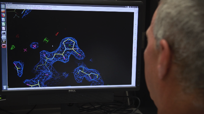
In order to better understand the center of human intelligence in the brain, Ruogu Fang uses artificial intelligence.
Fang, an assistant professor in the Biomedical Engineering Department, has used machine learning to understand, capitalize and model different aspects of brain dynamics for the last ten years. She works extensively on neuroimaging and keeps in mind the potential ways that AI can help advance brain studies.
Improving patients’ treatment
During computerized tomography (CT) scans, patients become exposed to harmful radiation. Because this method of discovering internal issues is widely popular, researchers like Fang are interested in reducing that level of health risk while still producing high quality images.
It is estimated that every 2000 CT scans will cause one future cancer, i.e., 40,000 cases of future cancers from 80 million CT scans every year. The radiation output of a CT brain perfusion exam is typically 4 to 10 times of that in a routine head CT exam. Fang’s team of researchers has dedicated ten years to working on AI and machine learning engines that can design proper deep learning framework for low-dose CT perfusion scan restoration (a.k.a. auto-ML).
As medical imaging in healthcare continues to serve as a diagnostic tool, Fang looks to use AI in response heterogeneity and personalized treatment of cognitive aging for conditions like Alzheimer’s disease.
“Transcranial direct current stimulation (tDCS) is a non-invasive brain stimulation, when paired with cognitive training, has shown promise to remediate age-related cognitive decline and alter trajectories toward Alzheimer’s disease.” Fang said, adding that they use machine learning to predict who will improve in cognitive functions after intervention. “We’re going to see whether this artificial intelligence method can help doctors or medical professionals decide who will respond.”
When patients react to brain stimulation through precision dosing, researchers like Fang can have a better look at what it takes to slow down or pause the progression of Alzheimer’s disease and other conditions by analyzing the current distribution in tDCS in the brain through multimodal neuroimaging.
Similarly, these scientists work with electroencephalogram (EEG) to predict treatment outcomes for chronic pain. Spinal cord stimulation (SCS) masks pain signals before reaching the brain by surgically placing the device under the patient’s skin, after which it will send mild electrical pulses to the spinal cord.
“It’s really challenging to forecast which patients will respond well because it doesn’t count for personal characteristics, like demographics information or physical state,” Fang said, explaining that the stimulation is meant to mask the communication from pain to brain. “Their EEG data is highly correlated with their pain and sensation perception and the signal from their lower body to their brain.”
Fang ultimately works to combine aspects of every project to fill in gray areas that connect brain functions through artificial intelligence.
“There is actually a pretty big gap (in communication) between the two fields of computer science and healthcare, and huge opportunities if we can stimulate the conversation between those two groups of people,” Fang said.
Dr. Ruogu Fang will be sharing more on her research projects during her virtual seminar with the Informatics Institute, Artificial Intelligence in Precision Brain Dynamics. RSVP here.
This story originally appeared on UF Informatics Institute.
Check out more stories on the UF A.I. Initiative.

